
Home Institution: St. Cloud State University
Major/Minor: Chemistry
MRSEC Mentor: Marc Hillmyer
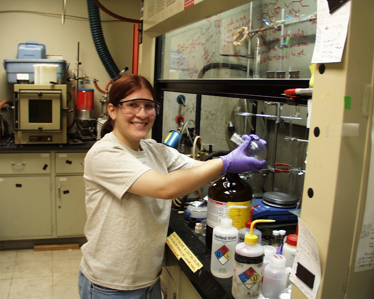
Synthesis of Fluorinated Oxadiazolines
Recently, much attention has been given to substituted _3Ð1,3,4Ðoxadiazolines as convenient sources for various types of carbenes. Of specific interest to the Hillmyer group at the University of Minnesota are fluorinated carbenes, for addition to unsaturated polymers. It is hoped that by controlling the amount of fluorine atoms attached to each repeat unit in the polymer, the solubility, as well as other physical characteristics, can be modified to those desired. In the course of this research, syntheses were developed for the generation of substituted _3 Ð 1,3,4 Ð oxadiazolines in general, and 2Ð(2,2,2Ðtrifluroethoxy)Ð2ÐmethoxyÐ5,5ÐdimethylÐ_3Ð1,3,4Ðoxadiazoline in specific. Syntheses of other fluoroalkoxy substituted _3Ð1,3,4Ðoxadiazolines were also attempted, with widely varying results. Attempted addition of methoxyÐ2,2,2Ðtrifluoroethoxy carbene to polyisoprene in a stainless steel vessel at 110o C, and under high nitrogen pressure, was unsuccessful. It is hoped that modification of this procedure will allow addition of the carbene to the polymer to occur.
Home Institution: Gustavus Adolphus College
Major/Minor: Chemistry
MRSEC Mentor: Frank Bates
Blends of Epoxy Resins with Epoxidized Isoprene/Butadiene Block Copolymer
A series of epoxidized isoprene/butadiene block copolymers (IxB) were synthesized from poly(1, 4-isoprene)-block-poly(1, 2-butadiene) (IB). A copolymer IB was epoxidized with in situ generated dimethyl dioxirane. The epoxidation reaction was selective for 1, 4-isoprene repeat units. The epoxidized 1, 4-isoprene had varying levels of epoxidation, ranging from 96 % down to 10%. The series of colpolymer IxB was then used to form copolymer/epoxy resin blends at 5 wt % copolymer IxB and 20 wt % copolymer IxB. The blends were composed of copolymer IxB and stoichiometric amounts of poly(Bisphenol A-co-epichlorohydrin) and methylene dianiline. These blends were visually observed at room temperature, after a 70 hour cure at 55 ¡ C, and after a one hour cure at 200 ¡C. It was observed that as the percent epoxidation of copolymer IxB in the blend increased, the copolymer miscibility with the epoxy resin increased, and the domain size decreased. DSC and IR was used to analyzed the cure of the blends, and they both showed evidence of the copolymer IxB epoxy groups reacting with the epoxy resin precursors. Further work will be done to characterize the microstructures in the blends, and to determine when and how copolymer IxB reacts with the epoxy precursors during the cure.

Home Institution: Gustavus Adolphus College
Major/Minor: Physics/Pre-Engineering
MRSEC Mentor: Jack Lewis
Cell Death Around Regions of Localized Neo-Cartilage Damage
Mechanical damage to articular cartilage is one factor that seems to be involved in the development of degenerative joint disease. Previous studies from our lab have documented cell death near cracks made due to mechanical trauma from impact loading, but minimal death near scalpel cuts. We have hypothesized that there would be a difference in the amount of cell death between areas of cartilage scratched by a relatively blunt object, such as a ball-point needle, and those cut by a scalpel. A fluorescent viability staining agent (Propidium Iodide) was used to quantify the amount of cell death around a scratch or cut in cultured cartilage tissue. Thresholding and measurement techniques incorporated into Metamorph image analysis software were used to relate the distance from damage to the relative amount of cell death. This comparison showed a vast difference between scratches, which had ten times the death near the scratch death before trailing off, and cuts, which showed no discernable difference in the amount of cell death between the cut area and background. These results suggest that localized tissue damage may directly induce cell death in cartilage.
Home Institution: College of St. Benedict
Major/Minor: Chemistry
MRSEC Mentor:
Brownian Motion of Semi-flexible Polymer Chains
The Brownian motion of semi-flexibly polymers in dilute solution was explored through computer simulation. The computer program, Large-scale Atomic/Molecular Massively Parallel Simulator (LAMMPS), was used to simulate the behavior of semi-flexible polymer chains in solutions and observe the systemÕs response to an initial step shear. The relaxation of the polymer chains from their initially deformed conformations was analyzed in terms of the stress in the system. The total stress in the system was decomposed into three components: stress due to the orientation of the molecules, stress due to the curvature of the molecules, and stress due to the tension in the bonds of the molecules. Calculations of the cross-correlations of these three components of the stress indicate a possible correlation between the orientation stress and the curvature stress, suggesting that modifications are necessary in the calculation of the curvature stress. The effects of altering system parameters were also explored. Reproducible results were obtained for the calculation of the tension stress while increasing or decreasing the damping constant, which correspondingly increased or decreased the diffusivity of the polymers in solution.

Home Institution: Carelton College
Major/Minor:
MRSEC Mentor: Michael Ward
Synthesis of Tetra(carboxy)TTF and Tris(4-pyridyl)benzene for the Purpose of Forming Redox-active Heterocrystals
The redox active tetra(carboxy)tetrathiafulvane was successfully synthesized and isolated using a coupling reaction between 4,5-dicarbomethoxy-1,3-dithiole-2-thione followed by a basification reaction using potassium hydroxide to convert the tetrathiafulvalene (TTF) tetra(methyl ester) into the TTF tetra(carboxy) acid. 1,3,5-tris(4-pyridyl)benzene was also synthesized using a multi-step procedure involving the formation of a tri(methyl)tinpyridine reagent which was then reacted with tribromobenzene. Column chromatography on an alumina gel column was then attempted in order to isolate the desired final product. This purification procedure proved unreliable, although some pure product was isolated. The procedure used to synthesize 1,3,5-tris(4-pyridyl)benzene should prove to be a general method capable of yielding numerous poly(4-pyridyl)-substituted benzenes. Efforts were also directed towards the synthesis of di(carboxy)TTF and ferrocinedialdehyde.
Home Institution: Carleton College
Major/Minor: Chemistry
MRSEC Mentor: Michael D. Ward
Flexible Extended Lenth Pillars for Open Framework Structures
Crystal engineering of guanidinium disulfonate networks has demonstrated two main types of host/guest lattice architecture. Work proceeded in an attempt to pinpoint the guest volume at which the architecture changed from bilayer to brick. In the [G]2[4,4-biphenyl disulfonate] guest clathrates, the bilayer isomer formed with guests of molecular volume less that 128 . The brick isomer is formed most often with guests larger than this value. Work was also done to synthesize and incorporate flexible extended length disulfonate pillars into these open framework structures. The longer flexible pillars, [G]2[1,2-bis(p-sulfophenoxy)ethane] [G]2[1,2-bis(p-sulfophenoxy)propane], were synthesized and successfully incorporated into host/guest clathrates. A sterically hindered pillar. [G]2[1.2-bis{3,5-dimethyl-4-sulfophenoxy)ethane}, was synthesized in hopes of creating a pillar that would be forced into the brick architecture due to sterics, but has yet to be incorporated into host/guest clathrate. Finally, the synthesis of a hetero pillar presented the possibility of a double bilyer structure, however, this too has to form a crystal structure.
Home Institution: University of Minnesota
Major/Minor: Physics
MRSEC Mentor: Dan Dahlberg
Frontiers of AFM and MFM
In studies of the coupling between ferromagnets and antiferromagnets (F/AF), most experiments measure some physical property of the F/AF coupling energy without knowledge of the location in the sample being probed, i.e., most studies use bulk probes which measure a signal from the entire sample such as vibrating sample magnetometer magnetization measurements. Although this certainly provides information on the F/AF coupling, it now appears that for a complete understanding of the phenomena, spatially localized studies must be made. For example, the field training effects which occur when the magnetization of the ferromagnet is reversed is most likely associated with either some type of irreversible domain wall in the ferromagnet or antiferromagnet or a rotation of both magnetization sublattices in smaller antiferromagnetic crystallites. In order to study the spatial variations in the F/AF coupling, we have begun magnetic force microscopy studies with in situ magnetic fields of the magnetic reversal in F/AF bilayers. These studies will allow us to determine nucleation sites of t he magnetization reversal and compare the domain structure in the ferromagnet upon repeated cycles of the reversal. This type of investigation will provide a microscopic picture of the coupling and how the grain size of the antiferromagnet controls the process. Most of the effort to date has been to develop a proficiency in scanning probe microscopy.
Home Institution: Hamline
Major/Minor: Chemistry
MRSEC Mentor: Andreas Stein
Adsorption of Perfume Oils
Crystal engineering of guanidinium disulfonate networks has demonstrated two main types of host/guest lattice architecture. Work proceeded in an attempt to pinpoint the guest volume at which the architecture changed from bilayer to brick. In the [G]2[4,4-biphenyl disulfonate] guest clathrates, the bilayer isomer formed with guests of molecular volume less that 128 . The brick isomer is formed most often with guests larger than this value. Work was also done to synthesize and incorporate flexible extended length disulfonate pillars into these open framework structures. The longer flexible pillars, [G]2[1,2-bis(p-sulfophenoxy)ethane] [G]2[1,2-bis(p-sulfophenoxy)propane], were synthesized and successfully incorporated into host/guest clathrates. A sterically hindered pillar. [G]2[1.2-bis{3,5-dimethyl-4-sulfophenoxy)ethane}, was synthesized in hopes of creating a pillar that would be forced into the brick architecture due to sterics, but has yet to be incorporated into host/guest clathrate. Finally, the synthesis of a hetero pillar presented the possibility of a double bilyer structure, however, this too has to form a crystal structure.
Home Institution: University of Minnesota
Major/Minor:
MRSEC Mentor: Robert T. Tranquillo
Identification of Myofibroblasts in Compacted Tissue Equivalents
During certain phases of wound healing, fibroblastic cells share characteristics of normal fibroblasts and smooth muscle cells. These cells are commonly referred to as myofibroblasts. An immunohistochemical protocol was developed to identify myofibroblasts in tissue equivalents - tissue cells dispersed in collagen or fibrin gels. An in vitro model is used to observe cell behavioral characteristics of myofibroblasts in tissue equivalents. The tissue equivalents compact when immersed in medium supplemented with serum, and wound fluid and growth factors can be added toto simulate a wound environment. It may be possible to confirm the presence of my fiibroblasts with accepted cell markers using immunohistochemistry. Fibroblasts were stained specifically for alpha-smooth muscle actin expression, a standard marker for myofibroblasts. While most cells were positive for alpha-smooth muscle actin, the stress fibers commonly associated with myofibroblasts were not clearly visible. Instead, we observed diffuse staining of the cytoplasm. Therefore, our protocol will be amended to include other myofibroblast markers such as desmin and vimentin, as well as other cytoskeletal proteins. Hoever, with the current protocol, it is possible to observe and quantify the morphology and alignment of cells in the gel. By identifying myofibroblasts in tissue equivalents, we hope to find how they may be implicated in wound contraction.
Home Institution: Xavier University
Major/Minor:
MRSEC Mentor: Allen Goldman
Investigation of Two Dimensional Superconductivity in Niobium (Nb) Doped Strontium Titanate (STO)
Superconductivity is a phenomenon that gives rise to special physical properties. One is zero resistivity. The study of two-dimensional superconductors is inspired by the fact that high temperature superconductors are made out of layered material. Studying one layer is a much simpler problem than studying many layers. Also, two-dimensional system physics is different from one-dimensional and three-dimensional systems in that only in the case of a two-dimensional system can one have an insulator or a superconductor but not a metal. Samples of niobium doped strontium titanate will be analyzed and fabricated from an insulating substrate where metal atoms are deposited on the substrate until the atoms form a disordered film that allows current to flow. When niobium is placed on strontium titanate and diffused in, there exists a region in the material where the concentration of niobium in strontium titanate is such that this portion of the materials is superconducting. A metal gate consisting of gold is placed on the back of the material. A voltage is placed across the two thin films to form a capacitor. Depending on the voltage, the concentration can be controlled; concentration is the number of charge carriers per unit area. Measurements will be made of Resistance vs. Temperature and Concentration vs. Depth. In the experiment involving superconductor-insulator transitions in the two-dimensional limit, results show that as thickness is increased, the substrate becomes more conductive and then eventually superconducting. Similar results are expected; as the gate voltage (voltage across the capacitor) is increased the niobium strontium titanate film will become insulating. This actually means that when the voltage across the capacitor is increased, the number of charge carriers available for conduction will decrease; thus the concentration or the number of charge carriers per unit area can be controlled. These two systems will be studied; this study will enable us to examine the underlying nature of the superconductor-insulator transition.
Home Institution: Clark University
Major/Minor:
MRSEC Mentor: Edgar Arriaga
Investigating methods for Testing the Toxicity of Doxorubicin
The primary purpose of this experiment was to investigate successful methods for testing the toxicity of doxorubicin. Doxorubicin (DOX) is a cytotoxic anti-cancer anthrocycline that is an essential drug used in the chemotherapeutic treatment of various types of cancer. During this study, the resistance of cells to doxorubicin, the pharmacological effects of doxorubicin in cells, and the maximum concentration of doxorubicin that cells can be exposed to without dying were investigated. One method that was examined for testing the toxicity of doxorubicin was Lethal Dosage at 50% survival (LD50). This value refers to the doxorubicin concentration that is able to enter and kill at least 50% of either type of cells (NS-land MAK). The LD50 value(s) (20µM DOX) was correlated with the fluorescence of cells which was viewed via microscopy. To test the efficiency of this method, a more reliable measurement of dead cells was taken by the Capillary Electrophoresis (CE-LIF). These results were compared as a standard as a standard to the results from the LD 50 technique. Once the drug was detected inside the cell, it was necessary to discover where the drug was localized inside the cell (which organelle). A further advancement in this research will include the use of the centrichip; a modern method of cell separation. Again, fluorescence will indicate which organelle is effected by the drug. The data obtained from this technique may be compared with data from the CE-LIF instrument in order to test the accuracy of the results. Moreover, it is suspected that the drug might appear in the nucleus, in the mitochondria, or in the cytoplasm of the cell. The cells are resistant to DOX at low concentrations and low incubation times. As shown in the data analysis, 5µM DOX is the maximum amount of the drug that can be used to treat cells without leaving them with severe cytotoxic effects. Additionally, it is expected that DOX appears in the nucleus because of the drug's ability to inhibit nucleic acid synthesis and mitotic activity which both occur in this organelle.
Home Institution: Clark University
Major/Minor:
MRSEC Mentor: Christopher Macosko
The Effects of Nucleating Agents on the Formation of Polystyrene Foams
Microcellular foams, which have cell diameters of 10 µm or less, are more advantageous than conventional foams because of improved tensile strength, improved fracture toughness, and eliminating the need for CFCs as blowing agents. Polystyrene is a polymer that is commonly foamed. Nucleating agents can be used in different combinations to control the number and size of cells necessary to create the desired microcellular foam. The purpose of this research is to determine what types and amounts of nucleating agent would be the most effective in forming polystyrene foam. In this research, CO2, and inert blowing agent, is used to form polystyrene foam with 0%, 0.5%, 1%, and 2% nucleating agents added to it. The nucleating agents used were carbon black, silica particles, and clay. Based on the data collected, it seems that adding nucleating agents to the polystyrene did not increase the number of bubbles nucleated, but instead decreased the number of bubbles. Polypropylene, which is difficult to foam, was also examined. The foaming difficulties are thought to be due to rheological properties, such as viscosity. Rheological measurements were taken of PP1, PP2, and three commercial polypropylenes with special emphasis on measurements of modulus and viscosity versus frequency. Results of PP 1 and PP 2 were compared to data received from Germany, while the three commercial polypropylenes were compared to each other. The PP 1 results matched the data from Germany, but the PP 2 results did not agree very well. The results from the three commercial polypropylenes were nearly identical.
Home Institution: North Carolina State University
Major/Minor:
MRSEC Mentor:
Home Institution: North Carolina State University
Major/Minor:
MRSEC Mentor:
Home Institution: Gustavus Adolphus College
MRSEC Mentor: Jack Lewis
The Effects of Bio-Active Glass on Rabbit Chondrocyte Development
Arising from the need produced by injury and arthritis, there is much interest in the in vitro creation of soft/hard tissue interfaces. Since bioactive glass has been shown to affect the differentiation of a variety of cell types, our project involved examining the effects of a bioactive glass on chondrocyte development. Cartilaginous tissues make a number of types of interactions with hard tissues such as bone, and can in addition be mineralized. The overall goals of this project were to assess whether bioactive glass beads had an effect on chondrocyte differentiation and whether or not contact with beads led to a tendency toward mineralization of either the beads or the tissue matrix. Initial experiments established that culture of rabbit chondrocytes with beads led to increased expression of alkaline phosphatase [a marker for mineralizing tissue] as assessed by cytostaining using an alkaline phosphatase specific stain. Biochemical analysis showed that at certain times [1-2 weeks] alkaline phosphatase activity was enhanced in cell lysates. Control experiments using silicates in the absence of beads indicated that contact with beads [rather than release of silicate from he beads] was necessary for this effect. Similar effects were observed with cow chondrocytes but the time scale for observed elevation of alkaline phosphatase was longer. Scanning electron microscope analysis of long term cultures [6 weeks] of chondrocytes, with bioactive glass beads added after two weeks suggested that the presence of the beads led to increased mineralization of the collagen matrix produced by the cells. Analysis of the beads [with and without prior removal of the organic matrix] by x ray analysis indicated that the organic matrix was the location of precipitated apatite crystals at short incubation times [2 week exposure to bioactive glass] but that with longer exposure apatite was deposited onto the glass as well as in the matrix. These results indicate that addition of bioactive glass to differentiating chondrocytes has an impact both on their differentiation and upon subsequent mineralization of the developing tissue.
Home Institution: St. Olaf College
MRSEC Mentor: Christopher Palmstrøm
The Effects of Bio-Active Glass on Rabbit Chondrocyte Development
Working directly with a team of graduate students, we assisted in the preparation of novel semiconductor samples with ferromagnetic contacts for spin injection devices. A range of characterization techniques including x-ray diffraction, Rutherford backscattering, and Atomic Force Microscopy. Magnetic property measurements relied on a Vibration Sample Magnatometer, Magnetic Force Microscopy, and the magneto-optic Kerr effect. Current-voltage and capacitance-voltage measurements of these diodes was used to determine the electric properties. Emphasis was placed on photolithography and diode construction, along with the construction and maintenance of ultra high vacuum (UHV) equipment in the lab. Short term projects have included construction an in situ (in vacuum) Schottky barrier height measuring system and vibration detection and isolation of key UHV equipment in preparation for installation of an in situ Scanning Tunneling Microscope Atomic Force Microscope device.
Home Institution: Sitting Bull College
MRSEC Mentor: Lorraine Francis
Interaction of Bioactive Glass with Collagen
Bioactive glasses in the Ca Mg phosphosilicate system were prepared and their bonding to collagen gels and demineralized dentin (largely collagen) was investigated. Bioactive glass beads were prepared by melting glass powder in a platinum crucible and pouring the melt onto an aluminum block. After annealing and polishing, the glass beads were placed adjacent collagen gels in vials containing artificial saliva. Type I collagen derived from calf skin was used to make the gel according to the protocol established by Knapp et al. (1997). The gel/glass combinations were held at 37¡C for at least 3 weeks. During the incubation period, the gel was compressed under the weight of the glass and migrated to cover the entire piece of glass. Upon removal, the pair was firmly bonded together and a white deposit was noted. In a separate experiment, glass beads were placed against pieces of dentin and demineralized dentin according the procedure developed by Efflandt et al. (1998). The pairs were secured with an elastic band and incubated for 3 weeks at 37¡C. Upon removal of the elastic band, the demineralized dentin/glass pair held together weakly while the dentin/glass pair was bonded more strongly. Collagen/glass and dentin/glass samples were then treated with glutaraldehyde to fix the collagen and then carefully dried. SEM and XRD analysis showed that the collagen/glass pair did not form any crystalline phase at the interface while the dentin/glass pairs did. Extensive shrinkage of the collagen and demineralized dentin hindered the analysis. A method to create a sol-gel derived bioactive glass was also established. Using metalorganic precursors, a solution was created with a composition identical to the bioactive glass. The solution gelled when exposed to a humid atmosphere. The gel was dried and then heated to remove the organic constituents. Thermal decomposition of these organics was challenging and more work needs to be done with thermal schedules and atmosphere to eliminate them. Sol-gel derived materials were incubated with collagen gels and bonding was observed.
Home Institution: Macalester College
MRSEC Mentor: Paul Crowell
Construction of an Ultrafast Terahertz Spectrometer
It is possible to generate ultra-short pulses of electromagnetic radiation with femtosecond lasers. These pulses can be used to perform pulsed spectroscopy at THz (1012Hz) frequencies with picosecond time resolution. This is an important development in condensed matter physics, because many of the fundamental excitations of solids occur at THz frequencies. Support from the MERSEC College Faculty-Undergraduate Research Team Fellowship has allowed us to construct an ultrafast THz spectrometer. We used the system to study THz emission from InAs in high magnetic field, and have shown that InAs is an efficient radiation source for further experiments. Further work is under way to understand and optimize the THz emission from InAs. The THz spectrometer will then be used to perform time-resolved spectroscopy on a variety of systems, including traditional semiconductor quantum wells and magnetic semiconductors. These types of systems will be the building blocks of the spin injection devices which are the goal of the Magnetic Heterostructures Initiative.

Home Institution: Northwestern College
MRSEC Mentor: Dan Dahlberg
Wide Temperature Range AC Susceptometer
An alternating current (AC) susceptometer was constructed. The device will measure the magnetic susceptibility of small (approximately 2 mm x 2 mm x 1 mm) samples from 4 K up to room temperature. The device was built to fit inside a glass dewar, and can be cooled with liquid nitrogen or liquid helium. Magnetic field coils were also constructed, so that the sample can be subjected to a steady magnetic field. A computer data acquisition system is used to collect the data from the susceptometer. The device was tested using well-known materials of high purity (Dysprosium Oxide and Gadolinium Oxide). It was used to obtain a new result on a two-layer thin film sample (15 Angstroms of Cobalt Oxide on top of 1000 Angstroms of Permalloy on a substrate). The sample was cooled from room temperature in a magnetic field. The magnetic field was then turned off and varied from 0 Gauss to 142 Gauss. A peak at approximately 22 Gauss was observed.
The University is funded through the National Science Foundation MRSEC Program, Award DMR-1420003
UMN MRSEC
435 Amundson Hall, 421 Washington Ave. SE, Minneapolis, MN, 55455
P: 612-626-0713 | F: 612-626-7805